solid teal background

Duplex ultrasound evaluation of a tortuous, recurrent varicose vein pre- and post-Varithena

solid grey background

Case Study Contributor

Ala Atieh, B.S, RVT
Lead Vascular Technologist, MIMIT Health- IL


“Due to the severity of the patient’s vein disease and history of recurrent varicose veins, the treating doctor and I decided Varithena would be the best option.”

Patient Characteristics

A 59-year-old male patient presented with a history of varicose veins in bilateral lower extremities since he was a teenager. He has had ulcers in bilateral malleoli of extremities however none active at the time of visit. Patient describes a pain score of 7 on a subjective scale from 0 (no pain) to 10 (most intense pain possible).

Patient’s associated pain and swelling symptoms are exacerbated with sitting and standing and affects his overall ability to work as a golf pro. Symptoms only partially controlled with compression therapy.

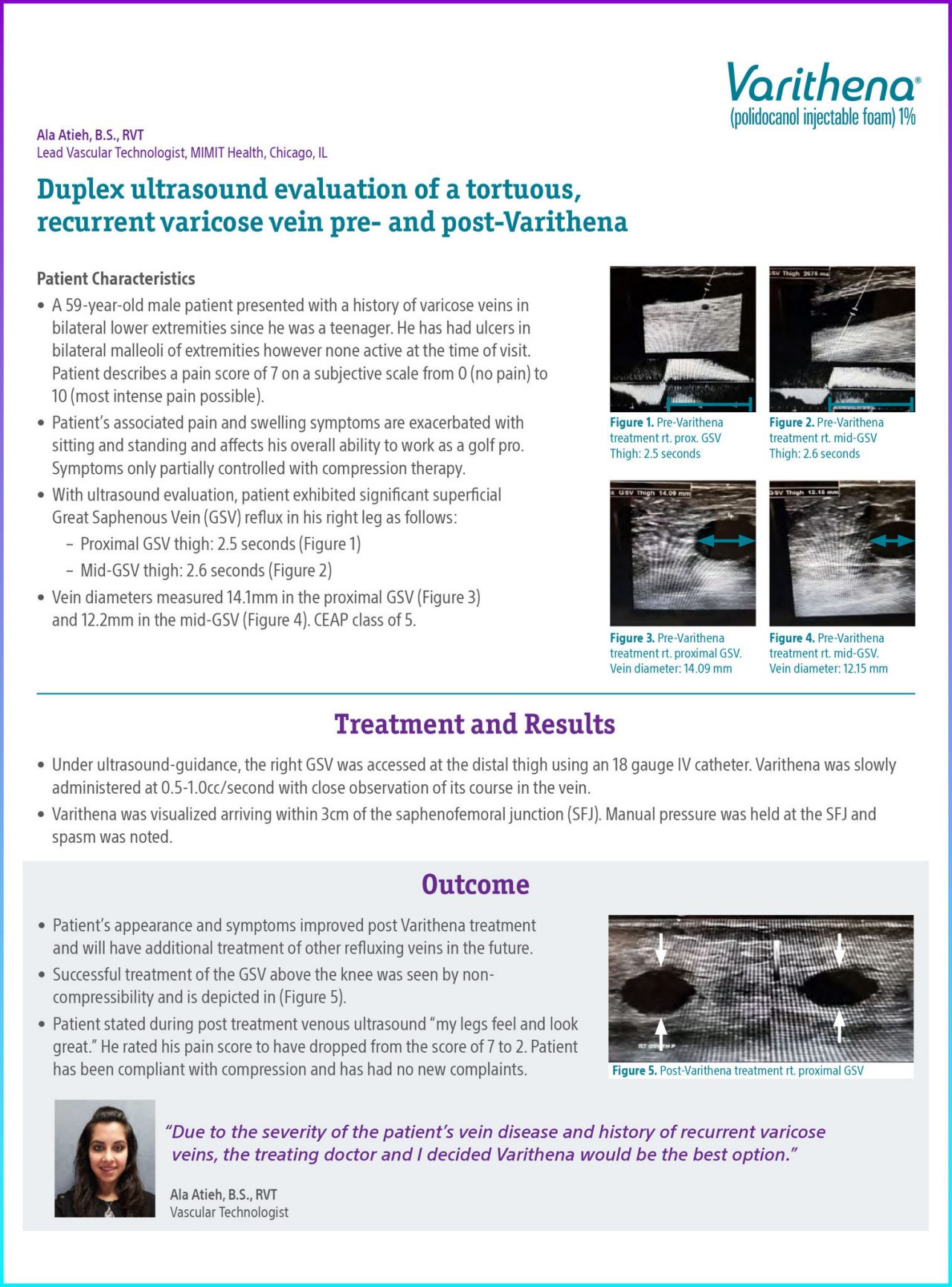
With ultrasound evaluation, patient exhibited significant superficial Great Saphenous Vein (GSV) reflux in his right leg as follows:

  • Proximal GSV thigh: 2.5 seconds (Figure 1)
  • Mid-GSV thigh: 2.6 seconds (Figure 2)

Vein diameters measured 14.1mm in the proximal GSV (Figure 3) and 12.2mm in the mid-GSV (Figure 4). CEAP class of 5.


Treatment and Results

Under ultrasound-guidance, the right GSV was accessed at the distal thigh using an 18 gauge IV catheter. Varithena was slowly administered at 0.5-1.0cc/second with close observation of its course in the vein.

Varithena was visualized arriving within 3cm of the saphenofemoral junction (SFJ). Manual pressure was held at the SFJ and spasm was noted.


Conclusion

Patient’s appearance and symptoms improved post Varithena treatment and will have additional treatment of other refluxing veins in the future.

Successful treatment of the GSV above the knee was seen by non-compressibility and is depicted in (Figure 5).

Patient stated during post treatment venous ultrasound “my legs feel and look great.” He rated his pain score to have dropped from the score of 7 to 2. Patient has been compliant with compression and has had no new complaints.


Documentation

Ultrasound of Varithena pre-treatment

Figure 1. Pre-Varithena Treatment Rt. Prox. GSV Thigh: 2.5 seconds

Ultrasound of Varithena pre-treatment

Figure 2. Pre-Varithena Treatment Rt. Mid-GSV Thigh: 2.6 seconds

Ultrasound of Varithena pre-treatment

Figure 3. Pre-Varithena Treatment Rt. Proximal GSV. Vein diameter: 14.09 mm

Ultrasound of Varithena pre-treatment

Figure 4. Pre-Varithena Treatment Rt. Mid-GSV. Vein diameter: 12.15 mm

Ultrasound of Varithena post-treatment

Figure 5. Post-Varithena Treatment Rt. Proximal GSV
Take this with you

Download case study

* Results from case studies are not necessarily predictive of results in other cases. Results in other cases may vary.

Indications

Varithena® (polidocanol injectable foam) is indicated for the treatment of incompetent great saphenous veins,  accessory saphenous veins and visible varicosities of the great saphenous vein (GSV) system above and below the knee. Varithena® improves the symptoms of superficial venous incompetence and the appearance of visible varicosities.

Important Safety Information

The use of Varithena® is contraindicated in patients with known allergy to polidocanol and those with acute thromboembolic disease. Severe allergic reactions have been reported following administration of liquid polidocanol, including anaphylactic reactions, some of them fatal. Observe patients for at least 10 minutes following injection and be prepared to treat anaphylaxis appropriately. Intra-arterial injection or extravasation of polidocanol can cause severe necrosis, ischemia or gangrene. Patients with underlying arterial disease may be at increased risk for tissue ischemia. If intra-arterial injection of polidocanol occurs, consult a vascular surgeon immediately.Varithena® can cause venous thrombosis. Follow administration instructions closely and monitor for signs of venous thrombosis after treatment. Patients with reduced mobility, history of deep vein thrombosis or pulmonary embolism, or recent (within 3 months) major surgery, prolonged hospitalization, or pregnancy are at increased risk for developing thrombosis. The most common adverse events observed were pain/discomfort in extremity, retained coagulum, injection site hematoma or pain, common femoral vein thrombus extension, superficial thrombophlebitis, and deep vein thrombosis.Physicians administering Varithena® must be experienced with venous procedures, possess a detailed working knowledge of the use of the duplex ultrasound in venous disease and be trained in the administration of Varithena®.

See Full Prescribing Information for Varithena®

Varithena™ is a registered trademark of Boston Scientific. All other trademarks are property of their respective owners.
PI-1263705-AA