blank pixel

Duplex ultrasound evaluation of a large refluxing branch of the GSV pre- and post-Varithena

    blank pixel

Case Study Contributor

Amanda C. Turner, RVT
Mount Auburn Hospital, Cambridge, MA


“After initial diagnostics and vein mapping, Varithena was considered the best alternative for this case. The patient had a previous history of RFA with recurrent tortuous varicosities post-ablation. The ablation was not able to close off the additional tributaries and the distal GSV in the calf. Foam allowed us to get into the smaller vessels.”

Patient Characteristics

A 68-year-old, otherwise healthy, male with lifelong severe bilateral varicose veins presented to the clinic with pain, heaviness, itching and foot discomfort.

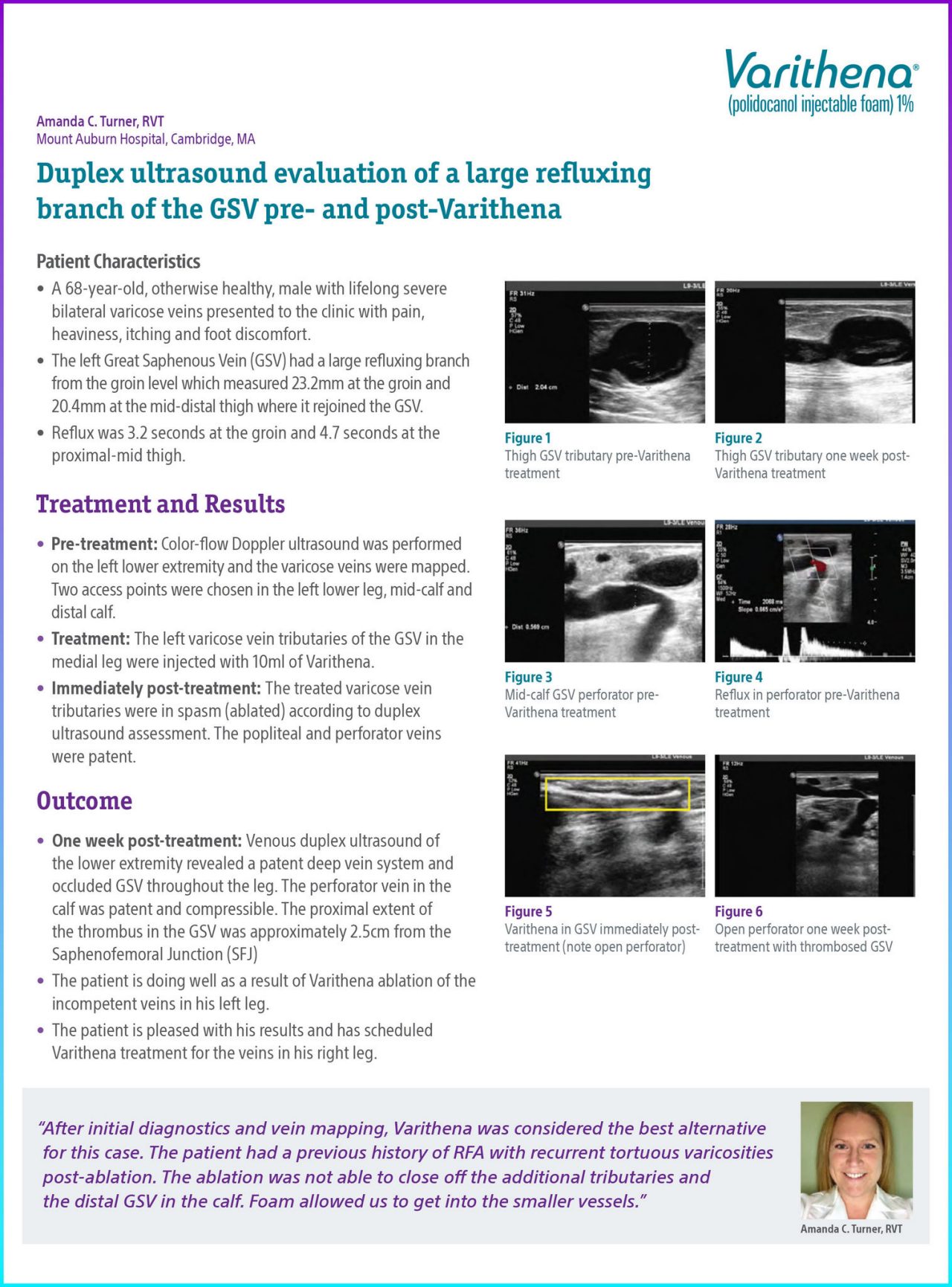
The left Great Saphenous Vein (GSV) had a large refluxing branch from the groin level which measured 23.2mm at the groin and 20.4mm at the mid-distal thigh where it rejoined the GSV.

Reflux was 3.2 seconds at the groin and 4.7 seconds at the proximal-mid thigh.


Treatment and Results

Pre-treatment: Color-flow Doppler ultrasound was performed on the left lower extremity and the varicose veins were mapped. Two access points were chosen in the left lower leg, mid-calf and distal calf.

Treatment: The left varicose vein tributaries of the GSV in the medial leg were injected with 10ml of Varithena.

Immediately post-treatment: The treated varicose vein tributaries were in spasm (ablated) according to duplex ultrasound assessment. The popliteal and perforator veins were patent.


Conclusion

One week post-treatment: Venous duplex ultrasound of the lower extremity revealed a patent deep vein system and occluded GSV throughout the leg. The perforator vein in the calf was patent and compressible. The proximal extent of the thrombus in the GSV was approximately 2.5cm from the Saphenofemoral Junction (SFJ).

The patient is doing well as a result of Varithena ablation of the incompetent veins in his left leg.

The patient is pleased with his results and has scheduled Varithena treatment for the veins in his right leg.


Documentation

Ultrasound of GSV Tributary pre-Varithena treatment

Figure 1. Thigh GSV Tributary pre-Varithena treatment.

Ultrasound of thigh with GSV tributary one week post- Varithena treatment

Figure 2. Thigh GSV Tributary one week post- Varithena treatment.

Mid-calf GSV perforator pre-Varithena treatment

Figure 3. Mid-calf GSV perforator pre-Varithena treatment.

Ultrasound of reflux in perforator pre-Varithena treatment

Figure 4. Reflux in perforator pre-Varithena treatment.

Varithena in GSV immediately post-treatment (note open perforator)

Figure 5. Varithena in GSV immediately post-treatment (note open perforator).

Open perforator one week post-treatment with thrombosed GSV

Figure 6. Open perforator one week post-treatment with thrombosed GSV.
Take this with you

Download case study

* Results from case studies are not necessarily predictive of results in other cases. Results in other cases may vary.

Indications

Varithena® (polidocanol injectable foam) is indicated for the treatment of incompetent great saphenous veins,  accessory saphenous veins and visible varicosities of the great saphenous vein (GSV) system above and below the knee. Varithena® improves the symptoms of superficial venous incompetence and the appearance of visible varicosities.

Important Safety Information

The use of Varithena® is contraindicated in patients with known allergy to polidocanol and those with acute thromboembolic disease. Severe allergic reactions have been reported following administration of liquid polidocanol, including anaphylactic reactions, some of them fatal. Observe patients for at least 10 minutes following injection and be prepared to treat anaphylaxis appropriately. Intra-arterial injection or extravasation of polidocanol can cause severe necrosis, ischemia or gangrene. Patients with underlying arterial disease may be at increased risk for tissue ischemia. If intra-arterial injection of polidocanol occurs, consult a vascular surgeon immediately.Varithena® can cause venous thrombosis. Follow administration instructions closely and monitor for signs of venous thrombosis after treatment. Patients with reduced mobility, history of deep vein thrombosis or pulmonary embolism, or recent (within 3 months) major surgery, prolonged hospitalization, or pregnancy are at increased risk for developing thrombosis. The most common adverse events observed were pain/discomfort in extremity, retained coagulum, injection site hematoma or pain, common femoral vein thrombus extension, superficial thrombophlebitis, and deep vein thrombosis.Physicians administering Varithena® must be experienced with venous procedures, possess a detailed working knowledge of the use of the duplex ultrasound in venous disease and be trained in the administration of Varithena®.

See Full Prescribing Information for Varithena®

Varithena™ is a registered trademark of Boston Scientific. All other trademarks are property of their respective owners.
PI-1263705-AA